Gray-scale morphology is a set of image processing techniques that operates on gray-scale images using mathematical morphological operations. It’s particularly useful for enhancing image details, removing noise, and preparing images for analysis by focusing on their structure. Unlike binary morphology (which is for binary images), gray-scale morphology considers the intensity of pixels, making it suitable for continuous-tone images.
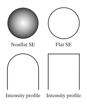


1. Structuring Element in Gray-Scale Morphology
In gray-scale morphology, a structuring element (SE) is a small matrix that defines the neighborhood over which morphological operations are performed. This element has values that represent pixel intensities, and it is used to probe and interact with the pixels in the image. Typical SEs are small matrices (e.g., 3×3, 5×5) that have various shapes such as a flat disk, square, or line.
2. Basic Operations in Gray-Scale Morphology
Gray-scale morphological operations include erosion, dilation, opening, and closing. Let’s break down each operation with its mathematical concept and an example.
A. Erosion (Minimization)
Definition: Gray-scale erosion of an image by a structuring element is the process of moving over each pixel in and replacing the pixel’s value with the minimum value covered by at that location.
Mathematical Concept:
where:
- is the gray-scale image.
- is the structuring element.
Example: Suppose we have a 3×3 region of an image:
If the structuring element is flat (all elements zero), erosion will take the minimum of each neighborhood. For a flat 3×3 SE, the output will be the minimum of all values in each 3×3 neighborhood around each pixel.
B. Dilation (Maximization)
Definition: Gray-scale dilation of an image by a structuring element is the process of moving over each pixel in and replacing the pixel’s value with the maximum value covered by at that location.
Mathematical Concept:
Example: Using the same 3×3 image from the previous example and a flat 3×3 SE, the output of dilation will be the maximum value in each 3×3 neighborhood around each pixel.
C. Opening
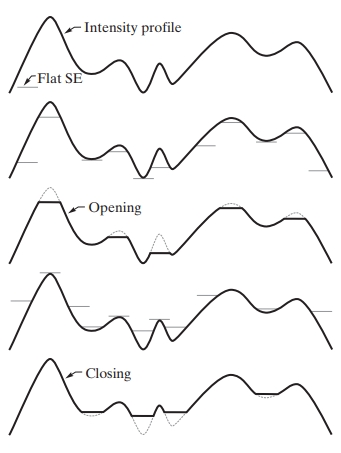
Definition: Opening is a two-step process: it first applies erosion, then applies dilation. Opening smooths out edges, removes small objects, and disconnects thin structures.
Mathematical Concept:
Example: For the image and a 3×3 SE, opening will erode , which reduces intensity values in bright regions, followed by dilation to restore some of the structure.
D. Closing
Definition: Closing is also a two-step process: it first applies dilation, then erosion. It is useful for closing small holes and connecting narrow breaks in an image.
Mathematical Concept:
Example: For the image , closing will first dilate , which enhances bright areas, followed by erosion to smooth out the result.
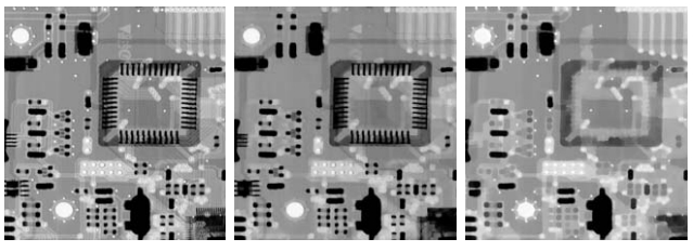
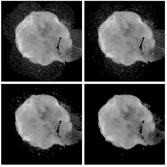
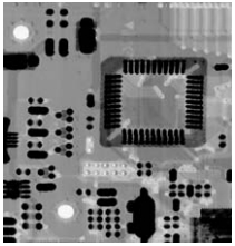
(b)–(d) Results of performing opening and closing sequences on the original image using disk-shaped structuring elements with radii of 1, 3, and 5 pixels, respectively.
(Original image courtesy of NASA.)
Image size: 566 x 566 pixels.
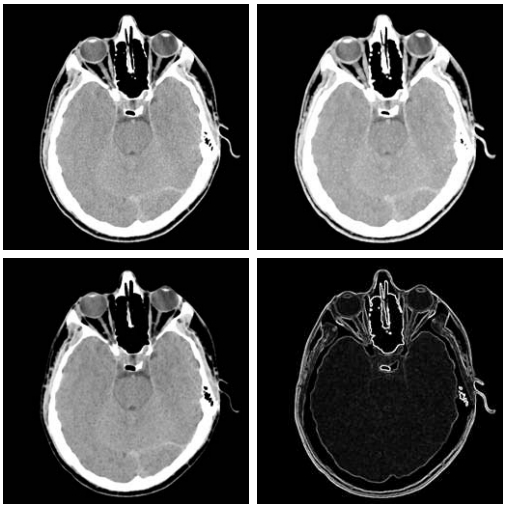
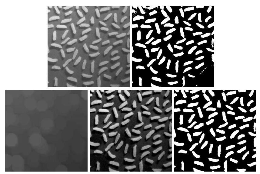
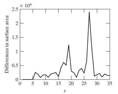
3. Some Basic Gray-Scale Morphological Algorithms
Gray-scale morphology can be applied to create specific algorithms useful in image processing.
- Top-Hat Transformation: Highlights small structures that are lighter than their surroundings.
- Bottom-Hat Transformation: Highlights small structures that are darker than their surroundings.
Example: Using the top-hat transformation, we can detect bright spots in a gray-scale image, which is particularly useful in astronomy to detect stars against a darker sky background.
4. Gray-Scale Morphological Reconstruction
Gray-scale morphological reconstruction is a technique that involves reconstructing an image by iteratively dilating a “marker” image until it fits within the boundaries of another “mask” image. Reconstruction is effective in removing undesired artifacts without distorting the shape of objects.
- Erosion-Based Reconstruction: It starts from a marker image and iteratively applies erosion until the marker conforms to the mask.
- Dilation-Based Reconstruction: It starts from a marker and applies dilation until it conforms to the mask.
Mathematical Concept: Let be the mask image and the marker image. The reconstruction of from by dilation, denoted , is obtained by:
Example: If we want to highlight regions that have bright cores in an image (e.g., cells in a microscopy image), we can use a gray-scale morphological reconstruction by dilation. The core regions (marker image) are iteratively dilated to match the boundary defined by the mask image (original image).
Summary of Operations
| Operation | Description | Mathematical Representation |
|---|---|---|
| Erosion | Reduces brightness in bright regions | |
| Dilation | Enhances brightness in bright regions | |
| Opening | Removes small bright areas | |
| Closing | Fills small dark areas | |
| Top-Hat | Detects small bright structures | |
| Bottom-Hat | Detects small dark structures | |
| Reconstruction | Recovers specific shapes in an image |
Each of these techniques is essential in preprocessing and analyzing gray-scale images, especially in fields like medical imaging, industrial inspection, and remote sensing, where structural details in continuous-tone images are key to analysis.
References
- Books:
- Gonzalez, R. C., & Woods, R. E. (2018). Digital Image Processing (4th Edition). Pearson.
- Haralick, R. M., & Shapiro, L. G. (1992). Computer and Robot Vision, Vol. 1. Addison-Wesley.
- Serra, J. (1983). Image Analysis and Mathematical Morphology. Academic Press. Jean Serra is one of the founders of mathematical morphology
- Research Articles:
- Vincent, L., & Soille, P. (1991). Watersheds in Digital Spaces: An Efficient Algorithm Based on Immersion Simulations. IEEE Transactions on Pattern Analysis and Machine Intelligence, 13(6), 583–598.
- Soille, P. (2003). Morphological Image Analysis
- Online Resources:
- MathWorks Documentation on Morphological Operations
- OpenCV Documentation on Morphological Operations
